Pediatric Urology › PUJ obstruction
PUJ Obstruction in Children: When Is Pyeloplasty Needed?
By Dr. Koushik Herle, MCh, DNB (Paediatric Surgery), Consultant Paediatric Surgeon and Paediatric Urologist, Manipal Hospital, Sarjapur–Marathahalli Road, Bengaluru. Medically reviewed: 22 October 2026
The pregnancy scan report says your baby’s kidney looks swollen. After birth, another scan mentions something called a PUJ obstruction. Your baby looks perfectly well, and you are not sure how worried to be.
PUJ obstruction is a narrowing where the kidney joins the tube that carries urine to the bladder. Urine drains slowly, so the kidney swells. It is one of the most common causes of kidney swelling (hydronephrosis) in children. Many mild cases need only regular scans. When the kidney is at risk, a planned operation called pyeloplasty fixes the narrowing.
Rest assured, PUJ obstruction is rarely an emergency. There is time to do the right tests and make a calm plan.
What is PUJ obstruction?
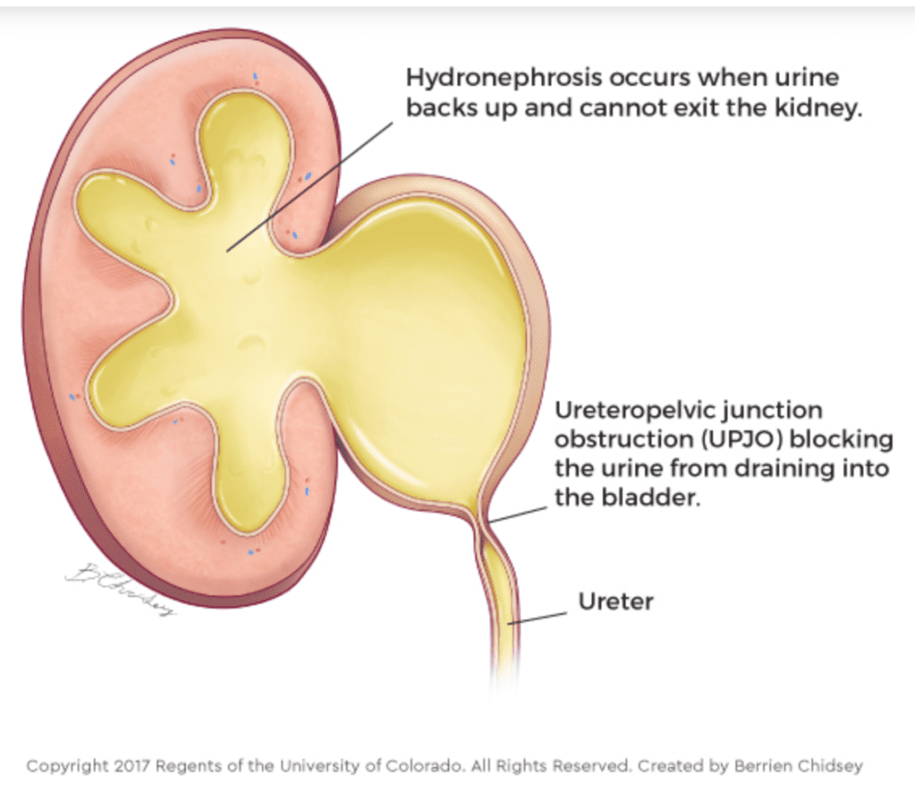
PUJ stands for pelviureteric junction. The pelvis of the kidney is the funnel through which urine leaves the kidney. The ureter is the tube that carries urine from the kidney to the bladder. The PUJ is the point where the two meet.
When this junction is narrow, urine cannot flow out freely. It backs up and stretches the kidney. Over time, this back pressure can slowly damage the kidney.
Why does it happen?
PUJ obstruction is usually congenital, which means the baby is born with it. Common reasons are:
- the muscle at the junction did not develop normally, so it does not pass urine along well,
- a blood vessel crosses the ureter and presses on it from outside,
- a narrow or kinked segment of the ureter itself.
Less often, it develops later in childhood, after infections, kidney stones or earlier surgery.
Signs parents notice
Many babies are picked up on a pregnancy scan that shows a swollen kidney. Read more on our page on antenatal hydronephrosis.
Please note: most PUJ obstructions cause no symptoms at all. Kidney function can be lost very slowly, over many years, without the child ever seeming unwell. This is why every baby whose pregnancy scan showed a swollen kidney must have follow-up scans after birth.
When symptoms do appear, they include:
- a swelling or lump in the tummy,
- repeated urine infections, often with fever and chills,
- pain in the side or back, sometimes with vomiting,
- blood in the urine,
- poor feeding or poor weight gain in babies.
Tests after birth
- Ultrasound: the first test. It shows how swollen the kidney is and is repeated over time.
- Diuretic renal scan (DTPA, MAG-3 or EC scan): a nuclear medicine scan. A small amount of tracer is injected and followed as it passes through the kidney into the urine. It shows how well each kidney works and how badly the junction is blocked. This test tells us whether surgery is needed now or not.
- Micturating cystourethrogram (MCU or VCUG): checks for urine flowing backwards from the bladder into the kidney (vesicoureteric reflux), which can occur along with PUJ obstruction.
- MRI urogram: used in some complex cases to see the anatomy in detail, including a crossing blood vessel.
Does every PUJ obstruction need surgery?
No. Many mild cases, especially in newborns with good kidney function, settle or stay stable. These children are watched with regular ultrasound scans and, when needed, a repeat renal scan.
Surgery is advised when:
- the function of the affected kidney is reduced,
- the swelling keeps getting worse on follow-up scans,
- the child has repeated urine infections,
- there is pain or blood in the urine.
If a truly blocked kidney is left untreated, it can lead to permanent kidney damage, repeated infections and high blood pressure. In severe cases, kidney failure can follow.
How pyeloplasty is done
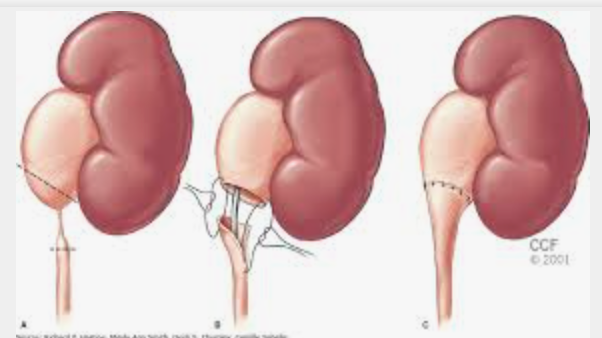
The standard operation is the Anderson-Hynes dismembered pyeloplasty. The narrow segment at the junction is removed, and the healthy ureter is joined back to the kidney pelvis in a wide, funnel shape so urine drains freely.
- Laparoscopic pyeloplasty: keyhole surgery through small cuts, using a camera. Recovery is usually quicker, with less pain and smaller scars. This is the approach most commonly used.
- Open pyeloplasty: done through a small cut on the side. This is preferred in very young infants, under about 3 months, with severe disease, in whom laparoscopy can be more difficult and risky.
Robot-assisted pyeloplasty: another keyhole option, in which the surgeon controls fine robotic instruments for extra precision. It is usually suited to older children. Robotic surgery is available at both Manipal Hospital Sarjapur Road and Manipal Hospital Electronic City.
How I decide
I look at three things: how well the kidney works on the renal scan, whether the swelling is getting worse over time, and whether the child has symptoms such as infection or pain. A stable kidney with good function is watched. A kidney that is losing function, or a child with symptoms, needs surgery.
Recovery after pyeloplasty
- Most children go home within 3 to 4 days after surgery.
- Pain is well controlled with regular medicines.
- A soft tube called a double J (DJ) stent is often placed inside the ureter during the operation. It keeps urine flowing while the join heals. It is removed after about 4 to 6 weeks, in a short day-care procedure.
- Follow-up scans check that the kidney is draining well and recovering.
Contact the surgical team the same day if: your child has a fever, burning or pain while passing urine, severe pain in the side, vomiting, or redness or discharge from the wound.
Will the kidney recover?
Surgery stops further damage. But kidney function that has already been lost may not fully come back. The nephrons, the tiny filters inside the kidney, do not grow back once they are destroyed. Partly injured nephrons can recover.
This is why timing matters. Finding PUJ obstruction early, and treating it when the scans say it is time, protects as much kidney function as possible.
The role of the paediatric surgeon
Paediatric urology is a major part of the MCh training in paediatric surgery. Paediatric surgeons are trained to assess and operate on conditions like PUJ obstruction in babies and children, using both laparoscopic and open techniques.
Questions parents ask
Is PUJ obstruction dangerous?
It is not usually life-threatening. But if a blocked kidney is not treated, it can be damaged permanently. Timely treatment protects the kidney.
Can PUJ obstruction go away on its own?
Mild cases can improve with time. Regular scans tell us whether the kidney is safe or whether surgery is needed.
Can surgery be done in small babies?
Yes. Pyeloplasty can be done safely in infants and even newborns when the kidney needs it.
Will my child need more than one operation?
In most children, one pyeloplasty is enough. The only planned second procedure is the short one to remove the DJ stent.
Will my child need long-term medicines?
Most children do not need any long-term medicines after a successful repair. Some babies are given a low-dose antibiotic while they are being watched, if advised.
Consult your paediatric surgeon early to understand your options. With regular scans and timely treatment, most children grow up with healthy kidneys.
Related pages
References
- European Association of Urology. EAU Guidelines on Paediatric Urology (chapter on dilatation of the upper urinary tract).
Clinic information
Dr. Koushik Herle, MCh, DNB (Paediatric Surgery), Consultant Paediatric and Neonatal Surgeon and Paediatric Urologist
Manipal Hospital, Sarjapur–Marathahalli Road, Bengaluru
Manipal Hospital, Electronic City, Bengaluru
Phone: +91 8217553785
This page is educational information and is not a substitute for individual medical advice. Drafted with AI assistance and medically reviewed by Dr. Koushik Herle.