Antenatal Hydronephrosis: Swollen Kidney on the Pregnancy Scan, Tests After Birth and Treatment
Medically reviewed by Dr. Koushik Herle, MCh, DNB (Paediatric Surgery), Consultant Pediatric Surgeon and Pediatric Urologist, on 20 November 2026
The anomaly scan report says your baby’s kidney is “dilated” or “swollen”. The pregnancy was going well, and now you have a new worry. Will the kidney be damaged? Will your baby need an operation?
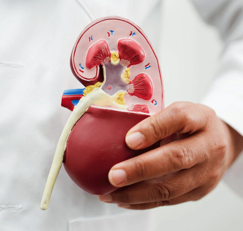
This finding is called antenatal hydronephrosis. “Hydro” means water and “nephros” means kidney. It means urine is collecting and stretching the kidney of the baby in the womb. It is one of the most common findings on the second-trimester anomaly (TIFFA) scan. The reassuring news: about 45 to 70 out of every 100 cases settle on their own. But every baby with this finding needs to be checked after birth.
Key points for parents
Antenatal hydronephrosis means a swollen kidney seen on the pregnancy scan, usually around 20 weeks.
About half or more settle on their own, but all babies need follow-up after birth.
The first scan after birth is best done after day 5, once feeding is well established.
Further tests, such as an MCU and a kidney (nuclear) scan, show whether there is reflux or a blockage.
Some causes stay silent for years while slowly harming the kidney, so follow-up matters even if the baby is well.
Any urine infection in a child under 2 needs a urine culture and an ultrasound before treatment.
What is hydronephrosis and hydroureteronephrosis?
Hydronephrosis means swelling of the kidney alone, because urine is collecting inside it. It is often caused by a narrowing where the kidney joins its drainage tube (the ureter). This is called PUJ obstruction.
Hydroureteronephrosis means swelling of both the kidney and the ureter (the tube that carries urine from the kidney to the bladder). It can be caused by urine flowing backwards from the bladder (vesicoureteric reflux, VUR), or by a blockage anywhere along the ureter, bladder or urine passage (urethra).
Think of it as a one-way road
Imagine the urinary system as a one-way road. The traffic (urine) should flow only from the kidney to the bladder.
Sometimes the road narrows, like a bottleneck, or has an obstacle. Traffic backs up and swells behind it. These are called obstructive causes.
Sometimes traffic tries to enter the one-way road from the wrong end. This also disrupts the flow and causes swelling. These are called reflux causes.
Causes
Some causes are temporary and settle by themselves. Others are structural problems that need treatment.
Blockage (obstructive) causes
Narrowing where the kidney meets the ureter: PUJ obstruction.
Narrowing of the ureter itself (ureteric stenosis).
Narrowing where the ureter enters the bladder: VUJ obstruction (megaureter).
Reflux causes
Primary vesicoureteric reflux, where the valve between the ureter and the bladder is weak.
Tip for parents: do not get the first ultrasound done on day one of life. A newborn is often a little dry in the first days, and the scan can make the kidney look normal when it is not. Do it after day 5, when feeding is well established and the baby is well hydrated.
Bring the pregnancy scan reports to the first visit. The grading of the swelling on those scans helps plan the follow-up after birth.
Symptoms: many children have none at first
The most important thing for parents to understand is that many of these conditions cause no symptoms at the start. They begin in the womb, but a PUJ obstruction, for example, can stay silent for years. VUR and VUJ obstruction often show up as repeated urine infections.
fever with no cough, cold, loose stools or other obvious cause;
poor weight gain;
anaemia (low blood count).
Any urine infection in a child under 2 years needs a urine culture and an ultrasound of the tummy. Giving antibiotics without these tests can hide the underlying problem, and it may go unnoticed until the kidney is damaged.
Routine screening of every baby is not needed. What matters is following up a swollen kidney seen on the pregnancy scan, investigating urine infections properly, and avoiding constipation, which helps keep the urinary system healthy.
Does it need treatment if there are no symptoms?
Often, yes. Blockages such as PUJ and VUJ obstruction can harm the kidney quietly, without any symptoms.
Think of the kidney as a hard-working filter. A blockage is like a roadblock. At first the kidney copes, but the pressure building up behind the block slowly damages it. This happens silently. Left untreated, the kidney may be badly damaged or stop working by the time the child reaches puberty. That is why a child with a significant blockage needs regular follow-up and treatment when the tests show the kidney is at risk, even if the child seems perfectly well.
Treatment
Mild swelling often settles on its own and needs only scans at intervals.
Moderate or severe swelling may need an operation to remove a blockage or correct reflux. This ranges from keyhole (minimally invasive) surgery, such as laparoscopic pyeloplasty, to open operations, depending on the cause.
Some babies are given a low daily dose of antibiotic while being followed up, to prevent infections.
Paediatric surgeons are trained in paediatric urology during their three-year MCh after general surgery, and guide the family through the treatment plan.
Long-term outlook
Most children with antenatal hydronephrosis grow up healthy with proper follow-up. Severe cases left untreated can lead to kidney scarring and poor kidney function. Keeping to the follow-up plan is the key.
Frequently asked questions
Will the swelling go away on its own?
In about 45 to 70 out of 100 babies, yes. But you can only know which group your baby is in by doing the scans after birth.
When should the first scan be done after birth?
Usually 5 to 7 days after birth. A scan on day one can be misleading because the baby may be slightly dry.
Does my baby need antibiotics?
Some babies do, especially those with reflux or marked swelling. Your doctor decides based on the scans.
Will my baby need an operation?
Most do not. Surgery is advised only when tests show a significant blockage, worsening swelling, falling kidney function or repeated infections.
Questions you may want to ask at your visit
How severe was the swelling on the pregnancy scan?
When should the first scan after birth be done?
Does my baby need an MCU or a kidney scan?
Should my baby take a daily antibiotic?
How often will follow-up be needed?
Consult your paediatric surgeon early to understand your options. Careful follow-up protects your child’s kidneys for life.
This page is educational information and is not a substitute for individual medical advice. Drafted with AI assistance and medically reviewed by Dr. Koushik Herle.