General and GI Surgery › Choledochal cyst
Choledochal Cyst in Children: Symptoms, Types and Surgery
By Dr. Koushik Herle, MCh, DNB (Paediatric Surgery), Consultant Paediatric Surgeon and Paediatric Urologist, Manipal Hospital, Sarjapur–Marathahalli Road, Bengaluru. Medically reviewed: 22 October 2026
Your child keeps getting tummy pain on the upper right side. You were told it is acidity. The medicines help for a while, and then the pain comes back. Sometimes the eyes look a little yellow. Then an ultrasound shows a swollen bile duct, and the report says choledochal cyst.
A choledochal cyst is a widening of the bile duct, the pipe that carries bile from the liver to the intestine. Children are born with it. It often hides behind vague, recurring tummy pain, jaundice that comes and goes, or unexplained pancreatitis, so it can be missed for months. Once it is found, a planned operation to remove it is usually the safest choice.
A typical story. A young child is treated more than once for gastritis because of recurring upper tummy pain. The pain keeps returning. An ultrasound finally shows a widened bile duct. After the cyst is removed, the pain goes away. This pattern is common, which is why a child with recurring tummy pain that does not fit the usual picture deserves an ultrasound.
What is a choledochal cyst?
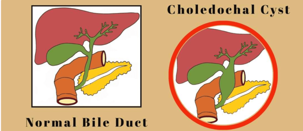
The word choledochal refers to the common bile duct. Bile is a digestive juice made by the liver. It flows down the bile duct into the small intestine, where it helps digest fat.
A choledochal cyst is not a separate sac of fluid, like a cyst in the ovary or kidney. It is the bile duct itself that balloons out at one or more places along its length, a little like a bulge in a hosepipe. You may also hear it called a bile duct cyst, biliary cyst or congenital bile duct dilatation. These all mean the same thing.
Most cases are found in infancy or early childhood. Some are now seen on a pregnancy scan before the baby is born. With better imaging, more cases are also found in older children.
Why does it happen?
The bile duct develops abnormally before birth. Nothing you did or did not do in pregnancy caused it. It is not linked to diet, stress or any medicine.
In many children, the pancreatic duct and the bile duct join too early, above their normal meeting point. Doctors call this an anomalous pancreaticobiliary junction. Digestive juice from the pancreas then flows back into the bile duct, irritates its wall, and makes it weak and wide over time. Not every child has this joint problem, so in some the duct wall itself may simply be weak.
Types of choledochal cyst
Surgeons use the Todani classification to describe where along the bile duct the widening is. The type matters, because it changes both the operation and the long-term outlook.
| Type | Where it occurs | How common |
|---|
| Type I | Widening of the common bile duct outside the liver | Most common, about 3 in 4 cases |
| Type II | A small pouch sticking out from the duct wall | Uncommon |
| Type III | A bulge where the duct enters the intestine (choledochocele) | Uncommon |
| Type IV | Several cysts, sometimes both inside and outside the liver | Second most common |
| Type V | Widening only of ducts inside the liver (Caroli disease) | Rare |
In children, Type I and Type IV make up the large majority, and both are treated by removing the cyst completely. Type III often behaves differently and can sometimes be treated through an endoscope instead of an operation. Type V is the most complex, because it can involve large parts of the liver.
Signs parents notice
Some children have no symptoms. The cyst is found on a scan done for another reason, or on a pregnancy scan. Others have symptoms because bile is not draining well. These differ with age.
| Babies | Older children |
|---|
| Jaundice that lasts or keeps returning | Pain in the upper right or middle of the tummy |
| Pale, clay-coloured stools | Jaundice that comes and goes |
| Dark urine | Nausea, vomiting, poor appetite |
| Poor feeding or poor weight gain | Itching of the skin |
| A lump felt in the tummy | Repeated pancreatitis or bile duct infection (cholangitis) |
Textbooks describe a classic triad of tummy pain, jaundice and a lump in the tummy. In real life, most children have only one or two of these. That is exactly why the diagnosis is often delayed.
Why it should not be left alone
When bile cannot flow freely, it backs up into the liver. An untreated choledochal cyst can lead to:
- cholangitis, an infection inside the bile ducts, with high fever and shivering,
- repeated pancreatitis,
- stones forming inside the widened duct,
- slow scarring of the liver and, in long-standing cases, cirrhosis,
- rupture of the cyst, which is uncommon but a surgical emergency,
- a higher long-term risk of cancer of the bile duct or gallbladder, especially if the cyst is left in place into adult life.
This cancer risk is the main reason the abnormal duct is removed completely, rather than simply drained or bypassed. Removal lowers the risk a great deal, but not to zero. That is why follow-up continues even after a good operation.
Tests your child may need
Blood tests show whether bile flow is affected. They include bilirubin, liver enzymes, infection markers if an infection is suspected, and amylase and lipase if pancreatitis is a concern. Blood tests alone cannot confirm a choledochal cyst.
Imaging confirms it:
- Ultrasound: usually the first test, and often enough to raise strong suspicion.
- MRCP (magnetic resonance cholangiopancreatography): a special MRI that maps the whole bile and pancreatic duct system without any endoscope. This is my preferred test for planning surgery in almost every case.
- CT scan: used occasionally to look at the surrounding anatomy or complications.
- ERCP: an endoscopic test that can show and treat some blockages. It is more invasive than MRCP, so I use it only in selected situations.
For most children, an ultrasound followed by an MRCP is all that is needed before surgery is planned.
Treatment: why surgery is usually the right choice
Parents often ask, can we just keep watching it? For most children, my honest answer is no. A choledochal cyst does not go away on its own, and the risks above build up the longer it stays. For Type I and Type IV cysts especially, I advise planned removal, ideally before repeated infections or liver damage set in.
The standard operation. For most Type I and Type IV cysts, the surgeon removes the diseased bile duct outside the liver, along with the gallbladder. Bile drainage is then rebuilt by joining the healthy bile duct directly to a loop of small intestine. This is called a Roux-en-Y hepaticojejunostomy. The name is long, but the idea is simple: remove the abnormal tissue completely, and build a clean, direct path for bile.
| Cyst type | Usual treatment |
|---|
| Type I and IV | Complete removal of the duct outside the liver, with Roux-en-Y hepaticojejunostomy |
| Type II | Removal of the pouch alone |
| Type III | Often treated through an endoscope if it causes blockage |
| Type V (Caroli disease) | Depends on how much of the liver is involved. It ranges from monitoring and treating infections to liver surgery or, rarely, transplantation, which is done at specialised liver centres |
Depending on the child’s age, the size of the cyst and any earlier inflammation, the operation is done as open surgery, by laparoscopy (keyhole surgery) or with robotic assistance. Robotic surgery is available at both Manipal Hospital Sarjapur Road and Manipal Hospital Electronic City. Keyhole and robotic surgery give smaller scars and often a quicker recovery. But the priority is always a safe and complete removal, and I choose the approach that serves that goal for each child.
Recovery after surgery
| When | What to expect |
|---|
| Day 1 to 2 | Pain controlled with medicines; sips of clear fluids started |
| Day 3 to 5 | Feeds increased as the bowel starts working; drain, if any, usually removed |
| Day 5 to 7 | Most children go home once they are eating well and comfortable |
| Week 2 to 4 | Gradual return to normal activity; wound check in clinic |
| From 3 months | Liver blood tests and an ultrasound to check that bile is draining well |
| Every year | Clinic review and scans to watch for narrowing at the join, stones or, rarely, cancer in the remaining bile duct |
Please keep every follow-up visit. The join can narrow months or years later, and a small long-term cancer risk remains in the leftover bile duct tissue. Missed follow-up is one of the most preventable reasons children come back with problems. Keep to the schedule even when your child looks and feels completely well.
Rest assured, with timely and complete surgery, most children go on to live normal lives, with no special diet and no recurring symptoms.
When to seek urgent care
Go to the emergency department the same day if your child has: fever with tummy pain, new or worsening yellow skin or eyes, severe or repeated vomiting, sudden severe tummy pain, or unusual drowsiness, confusion or weakness.
These signs are not specific to choledochal cysts. But in a child with a known or suspected cyst, they need same-day assessment, not waiting at home.
Questions parents ask
Is a choledochal cyst the same as a normal cyst?
No. It is a widening of the bile duct itself, not a separate fluid-filled sac.
Did I do something in pregnancy to cause this?
No. It is a condition the baby is born with. It has no known link to anything a mother ate, took or did in pregnancy.
Can we avoid surgery and just watch the cyst?
For most Type I and Type IV cysts, watching alone carries a real and growing risk of infection, pancreatitis and, in the long term, cancer. Planned removal is usually the safer path.
Will my child need follow-up for life?
Regular review is advised for the long term, even though most children feel completely normal within weeks of surgery.
Can it be done by keyhole or robotic surgery?
Often, yes. It depends on the child’s anatomy and the surgeon’s assessment. The aim is always a complete and safe removal.
Consult your paediatric surgeon early to understand your options. Finding and treating a choledochal cyst early protects your child’s liver for the long term.
Related pages
References
- Todani T, et al. Congenital bile duct cysts: classification, operative procedures, and review of thirty-seven cases including cancer arising from choledochal cyst. Am J Surg. 1977;134(2):263-269. PubMed.
Clinic information
Dr. Koushik Herle, MCh, DNB (Paediatric Surgery), Consultant Paediatric and Neonatal Surgeon and Paediatric Urologist
Manipal Hospital, Sarjapur–Marathahalli Road, Bengaluru
Manipal Hospital, Electronic City, Bengaluru
Phone: +91 8217553785
This page is educational information and is not a substitute for individual medical advice. Drafted with AI assistance and medically reviewed by Dr. Koushik Herle.