Medically reviewed by Dr. Koushik Herle, MCh, DNB (Paediatric Surgery), Consultant Pediatric Surgeon and Pediatric Urologist, on 20 November 2026
Your child keeps getting fevers. Each time it is a urine infection. Each time the antibiotics work, and a few weeks later the fever is back. Sometimes the hidden reason is a kidney stone.
This article uses one anonymised educational case to explain how kidney stones form in very young children, how they are found, why antibiotics alone may not be enough, and the treatment options: ESWL, RIRS, ureteroscopy and PCNL. For the full guide to kidney stones in children, see kidney stones in children.
Key points for parents
- Kidney stones can occur even in toddlers and babies.
- A stone can hide germs inside it, so urine infections keep coming back after every course of antibiotics.
- More than two proven urine infections in a young child need a kidney ultrasound to look for a stone or another cause.
- A stone that moves and blocks the kidney is an emergency. A thin tube called a DJ stent relieves the blockage.
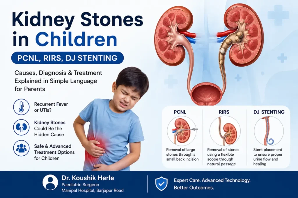
- Treatment depends on the stone’s size and position and the child’s age. Options include shock waves (ESWL), a telescope from below (RIRS) and a small puncture in the back (PCNL).
- Complete removal of every fragment matters, because left-behind pieces can restart infection.
An educational case
A boy of 20 months had a high fever. His doctors found a urine infection and treated it with a strong antibiotic for seven days. He got better. Three months later the fever returned, and then again. In a few months he had three separate urine infections, all with the same germ (E. coli). Each was treated with the right antibiotics, and each time he relapsed.
Tests were then done to look for a structural cause:
- Ultrasound showed two stones in the right kidney, of 1.2 cm and 0.5 cm, with mild kidney swelling.
- A bladder X-ray (MCU) was normal, with no urine flowing back up to the kidney.
- A nuclear kidney scan showed the kidney was draining and working adequately.
- Blood tests for calcium, uric acid, kidney function, parathyroid hormone and vitamin D were normal, so no metabolic cause was found.
How do kidney stones form in young children?
Urine contains dissolved minerals such as calcium, oxalate, uric acid and phosphate. Normally they stay dissolved. If they become too concentrated, they form crystals that stick together and grow into a stone. Doctors call this nephrolithiasis.
In this child, the most likely cause was an infection stone. The first infection probably did not fully clear deep inside the kidney. Germs can hide in a protective layer called a biofilm. Germs, pus and minerals then built up layer by layer, like a pearl forming around a grain of sand in an oyster. Over about three months, this grew into a 1.2 cm stone.
This is why antibiotics alone could not cure him. Each course killed germs in the urine, but germs inside the stone survived and started the next infection. The only way to break the cycle was to remove the stone completely.
For parents: if your young child has more than two proven urine infections, ask for a kidney ultrasound. Do not assume it is just “low immunity”. A stone is one of the important, treatable causes of repeated urine infections. See urine infections in children.
The emergency: when the stone moved
While tests were being done to plan surgery, the larger stone slipped out of its place and got stuck where the kidney joins its drainage tube (the pelvi-ureteric junction). This is one of the narrowest points in the urinary system. It was like a cork stuck in the neck of a bottle. Urine could no longer drain from the kidney. The child developed a swollen kidney, colicky tummy pain, vomiting and fever.
A child with fever, vomiting and severe tummy or side pain needs urgent care. Do not wait. Go to the nearest emergency department.
DJ stent: relieving the blockage
Under general anaesthesia, a thin telescope was passed into the bladder (cystoscopy), and a soft, thin tube called a DJ stent was passed up past the stone into the kidney. It is called “double-J” because it curls at both ends, like the letter J. Think of a thin drinking straw with curled ends, one inside the kidney and one inside the bladder, keeping the path open. It sits fully inside the body, with no outside wound.
As soon as it was in place, thick infected urine with pus began to drain. Over 48 hours the urine cleared. Antibiotics chosen from the urine culture were continued for nine days. Surgery to remove the stones was done only on day ten, once the infection had settled.
Operating on an infected, blocked kidney is dangerous, because it can spread germs into the blood (sepsis). That is why the infection is cleared first.
Treatment options for kidney stones in children
Stone surgery in children is not one-size-fits-all. The right choice depends on the stone’s size, position and type, the child’s age and anatomy, and whether there is infection or blockage.
1. Watching, with medicine to help the stone pass
Small stones, usually under 5 mm, can pass by themselves, especially if they are low down near the bladder. Plenty of fluids and sometimes a medicine that relaxes the drainage tube help. This is only for small stones with no blockage and no infection.
2. ESWL (shock wave lithotripsy)
Shock waves are aimed at the stone from outside the body, breaking it into fragments that pass in the urine. Nothing is put inside the body.
- Best for: stones under 1 to 1.5 cm in the kidney or upper tube, with no infection, in children over 1 year.
- Limits: works less well for hard, large or lower-kidney stones, may need several sessions, and cannot be used with infection or blockage.
In this case, ESWL was not suitable: the stone was larger than 1 cm, there was infection, and the stone had caused a blockage.
3. Ureteroscopy and RIRS (a telescope from below)
A thin telescope is passed through the urine passage and up the drainage tube. In RIRS, a flexible telescope goes all the way into the kidney, and a laser breaks the stone into dust. There is no cut on the skin.
- Rigid ureteroscopy is used for stones in the tube, but the tube of a very young child is very narrow and can be injured.
- RIRS works very well in older children and in selected younger ones, often after a stent has gently widened the tube first. Published stone-free rates are about 83 to 90 in 100 for stones up to 2.5 cm.
In this 20-month-old child, RIRS was harder for two reasons: the drainage tube was very narrow, and the stone was now loose in the open centre of the kidney, where it could roll away from the laser.
4. PCNL (a small puncture in the back)
PCNL stands for percutaneous nephrolithotomy: “percutaneous” means through the skin, “nephro” means kidney and “lithotomy” means stone removal. A narrow track is made from the skin of the back straight into the kidney. A thin telescope is passed through it, and the stone is broken up and removed. The wound is a small puncture of about 1 to 2 cm.
For children, smaller versions are used:
| Type | Track size | Used for |
|---|---|---|
| Standard PCNL | More than 7 mm | Very large stones. Rarely used in small children. |
| Mini PCNL | About 5 to 7 mm | Stones over 1 cm in children. The most common choice. |
| Micro or ultra-mini PCNL | Less than 5 mm | Babies and small children. |
- Best for: stones over 1 to 1.5 cm, lower-kidney stones, infection stones that need complete clearance and washing of the kidney, and very young children where RIRS access is difficult.
- Published stone-free rates: about 83 to 90 in 100 in children, including under 2 years, in experienced hands.
How the mini PCNL was done in this case
- Position: under general anaesthesia, the child lay face down, giving direct access to the back of the kidney.
- Puncture: using live X-ray guidance, a fine needle was passed through the skin of the back into the best part of the kidney to reach the stone.
- Widening the track: a guidewire was passed in and the track gently widened to fit the small telescope.
- Removing the stones: the larger stone was broken up and removed, and the smaller one was lifted out with fine forceps.
- Washing the kidney: the kidney was rinsed with sterile salt water until clear, removing pus and debris from months of infection.
- New stent and closure: a new DJ stent was placed from above, and the small wound closed with a stitch.
Both stones were fully cleared and the kidney washed clean, removing the source of the repeated infections.
Recovery and follow-up
- Hospital stay: usually 2 to 4 days after PCNL. Feeding usually restarts the next day.
- The DJ stent is removed 4 to 6 weeks later in a short procedure under brief anaesthesia. An ultrasound then checks the kidney is draining and no stone is left.
- Prevention: plenty of fluids, prompt and complete treatment of any future urine infection guided by a urine culture, and regular ultrasound and urine checks.
Infection stones come back in up to 85 out of 100 children if any fragments are left behind. Complete clearance is therefore very important.
| Procedure | Hospital stay | Back to activity | Stent removal |
|---|---|---|---|
| ESWL | Day procedure | Next day | Not usually needed |
| RIRS or flexible ureteroscopy | 1 to 2 days | 2 to 3 days | 4 to 6 weeks later |
| Mini PCNL | 2 to 4 days | 1 to 2 weeks | 4 to 6 weeks later |
| Emergency DJ stent | 1 to 2 days | Same day | At the stone operation |
Signs of a kidney stone in a child
- Fevers that keep coming back with no clear cause.
- Repeated urine infections, especially with the same germ each time.
- Colicky tummy or side pain. Young children may just be very irritable, scream or draw up their legs.
- Blood in the urine, which may look red or pink or show only on a urine test.
- Difficulty passing urine, or a weak stream.
- Vomiting with tummy or side pain.
- Poor weight gain.
Repeated urine infections in young children are never normal. More than two proven infections in a year, especially with fever, need a full check of the urinary tract.
Frequently asked questions
My 18-month-old has just had a second urine infection. Should I worry about a stone?
Two proven infections in a child under two should be investigated, starting with a kidney ultrasound. If a stone is seen, further tests help plan treatment.
Can E. coli cause a kidney stone?
Infection stones are classically linked to other germs such as Proteus and Klebsiella. But E. coli can form a biofilm, and incompletely cleared pus and debris can act as a base on which minerals build up.
Can a stone pass on its own?
Stones under about 5 mm may pass. Stones over 1 cm rarely do. A stone that holds germs must be removed completely, because antibiotics cannot clear the infection while it is there.
Is RIRS or PCNL better?
Neither is better for everyone. RIRS needs no cut and suits older children and selected younger ones. PCNL needs a small puncture but gives direct access, and suits larger stones, infection stones that need washing out, and very young children. Both have similar success rates in the right child.
Is PCNL safe in a child under 2?
Published studies show mini PCNL is safe and effective in children under 2, including babies, with careful selection, small instruments, an experienced paediatric anaesthesia team, and infection cleared before surgery.
Does a DJ stent hurt?
It is placed and removed under anaesthesia. While it is in, older children may feel they need to pass urine more often or feel mild discomfort. Young children usually tolerate it well.
Will the stone come back?
It depends on the stone type. Complete removal, plenty of fluids, prompt treatment of infections and regular checks greatly lower the risk. If a metabolic cause is found, diet changes and medicines help.
Questions you may want to ask at your visit
- How big is the stone, and where is it?
- Is there infection or blockage now?
- Which treatment do you advise for my child’s age and stone, and why?
- Will my child need a DJ stent, and when will it come out?
- What tests are needed to find the cause?
Consult your paediatric surgeon early to understand your options. Finding and removing a hidden stone can end the cycle of repeated infections.
Related pages
- Kidney stones in children
- Urine infections in children
- Pediatric urology
- Keyhole surgery for children
This article is educational information and is not a substitute for individual medical advice. Patient details are anonymised and shared for education. Drafted with AI assistance and medically reviewed by Dr. Koushik Herle (MCh, DNB Paediatric Surgery; KMC registration 90606).