In the media · The Times of India · Health · 4 July 2026
20-month-old boy’s persistent UTIs reveal a rare genetic disease that threatened his kidney
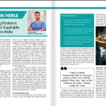
The Times of India reported on a toddler of 20 months whose repeated urinary infections turned out to be cystinuria, an inherited condition that forms kidney stones. A large stone had moved and blocked the flow of urine. The team placed a DJ stent as an emergency step, then cleared the stone with a keyhole laser procedure (mini-PCNL). He was feeding again within a day and went home after a short stay. Dr Koushik Herle, Consultant Paediatric Surgeon, is quoted on the precision that surgery in children under two demands.
Related: UTI in children: symptoms, tests and treatment
By Dr. Koushik Herle, MCh, DNB
Consultant Pediatric & Neonatal Surgeon | Pediatric Urology, Oncology & Minimally Invasive Surgery Manipal Hospital, Sarjapur–Marathahalli Road, Bengaluru
A 20-month-old boy from West Bengal had been suffering from recurrent urinary tract infections for almost six months, leaving his family desperately looking for answers. Initially suspected to have recurrent UTIs due to conditions like vesicoureteric reflux (VUR) or incompletely treated UTIs, the toddler was eventually diagnosed with a rare kidney stone problem. Seeking specialized care, the family traveled to Bengaluru, where doctors at Manipal Hospital Sarjapur Road successfully treated the condition through a minimally invasive procedure, saving the child from major open surgery.
The child’s symptoms had persisted for about half a year despite several courses of treatment. Further investigations revealed two stones in his right kidney, including a large one measuring 1.2 cm. Because of the patient’s unusually young age, the family sought advanced intervention in Bengaluru.
During the evaluation process, the situation became critical when the larger stone unexpectedly migrated from the kidney and lodged at the pelvi-ureteric junction (PUJ), completely obstructing urine flow. The child rapidly developed a high fever, vomiting, and severe abdominal pain due to the infected, obstructed kidney, necessitating emergency intervention.
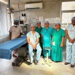
Dr Koushik Herle, Consultant Paediatric Surgeon, led a multidisciplinary team, including the hospital’s specialized Urology department, to perform an emergency DJ stenting procedure. This immediately relieved the obstruction and drained the infected urine. Once the patient was stabilized and treated with a course of culture-directed IV antibiotics, the medical team performed a highly specialized, minimally invasive procedure known as Mini Percutaneous Nephrolithotomy (Mini PCNL). Using advanced laser technology through a tiny puncture access point, the surgeons fragmented and removed the larger stone, cleared out the smaller stone, and thoroughly flushed the infected debris from the kidney. The entire procedure achieved total clearance without requiring a traditional surgical incision.
“Operating on an infant under two years old requires an extraordinary level of precision, as their delicate anatomy demands specialized paediatric instruments and a highly experienced multidisciplinary team. By opting for the Mini PCNL approach, we avoided the severe physical toll and extensive recovery associated with open surgery, ensuring the child could heal quickly with minimal scarring,” said Dr Koushik Herle, Consultant Paediatric Surgeon, Manipal Hospital Sarjapur Road.
A subsequent biochemical analysis revealed that the removed object was a rare cystine stone, which is typically linked to a genetic metabolic disorder called cystinuria. The medical team has recommended further genetic and metabolic screening to identify the underlying cause and establish a preventative plan against future recurrences.
The young patient recovered remarkably well after the procedure. He resumed feeding within 24 hours and was discharged following a brief hospital stay. Today, the child is active, healthy, and awaiting routine stent removal, with no visible marks left behind other than a tiny puncture wound on his back.
The young patient recovered remarkably well after the procedure. He resumed feeding within 24 hours and was discharged following a brief hospital stay. Today, the child is active, healthy, and awaiting routine stent removal, with no visible marks left behind other than a tiny puncture wound on his back.
“When a stone migrates and blocks urine flow, it creates an acute medical emergency that threatens kidney function. Our primary objective with the Mini PCNL technique was to eliminate the blockage and clear the infected debris as safely as possible. This advanced endourological approach successfully protected the child’s renal health, significantly mitigated post-operative pain, and allowed for an excellent cosmetic and clinical outcome,” said Dr Manohar Bhadrappa, Senior Consultant – Chief of Urology, Renal Transplant Surgeon & Uro-Oncology Surgeon, Manipal Hospital Sarjapur Road.
Kidney stones in infants and toddlers are exceedingly rare and are typically triggered by underlying metabolic conditions, genetic factors, or chronic infections. Early diagnosis and prompt therapeutic intervention are vital to safeguard young patients from permanent kidney damage and long-term complications. As demonstrated in this case, modern minimally invasive techniques like Mini PCNL offer a highly effective, low-pain alternative that ensures brief hospital stays and rapid recoveries for even the youngest patients.